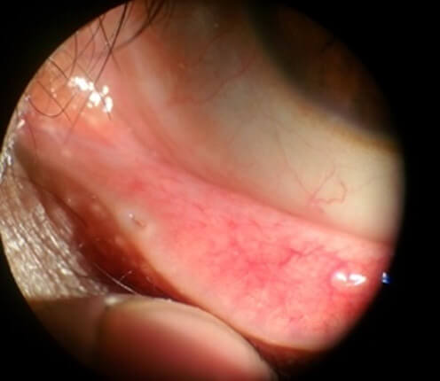
You may be one of many people who experience constant watery eyes or tearing. This excessive tearing may be secondary to dry eye disease, chronic irritation of the ocular surface, or a tear duct obstruction. When the latter is the culprit, the condition is called epiphora.
The tear duct system begins in the eyelids and runs down to the nose. Epiphora may be due to an obstruction of the upper, i.e., close to the eye, or lower tear duct. Obstructions of the lower tear duct are covered elsewhere in our blog (see blog). In this blog, we will discuss how to treat obstructions of the upper tear duct. Eyelids contain tiny openings on their inner surface through which the tears drain. These are called the lacrimal puncta (see figure).

A thin layer of mucosa lines the puncta. Several conditions may cause the mucosa to thicken or become swollen, causing the tiny puncta to close or narrow. This condition is known as punctal stenosis. When the caliber of the punctum decreases, the drainage of tears is significantly restricted, and patients will suffer from constant tearing. Fortunately, a minor surgical procedure can reverse these obstructions by reestablishing the patency of the punctum, thus, improving symptoms almost immediately.
A punctoplasty is the procedure of choice to treat obstructions of the lacrimal puncta. The operation is performed under local anesthesia in your doctor’s office. First, local anesthesia is injected into the area around the punctum. A blunt surgical instrument called a punctum dilator then dilates the punctum. Microsurgical scissors are used to make three incisions along the punctum to enlarge it. Finally, a special probe is introduced into the tear duct to confirm the patency of the whole system.
This procedure takes about 10 minutes and has a very short recovery. You will be instructed to apply eye drops three times a day for one week and come in for a follow-up appointment one week after the surgery. This operation has a high success rate, as long as there are no abnormalities or obstructions in the lower tear drainage. We have uploaded a short video to illustrate the procedure.


 |
|